
Bone Regeneration · 3D-BONE
3D-BONE: Customised Synthetic Bone Graft
The new digital frontier in reconstructive bone surgery. 3D-BONE offers preformed, synthetic bone grafts anatomically modelled with utmost precision to perfectly fit your patient's specific bone defect, drastically reducing intraoperative time.
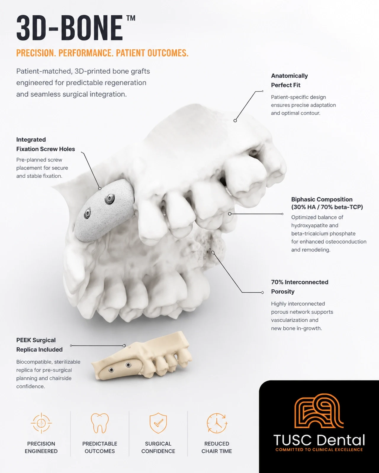
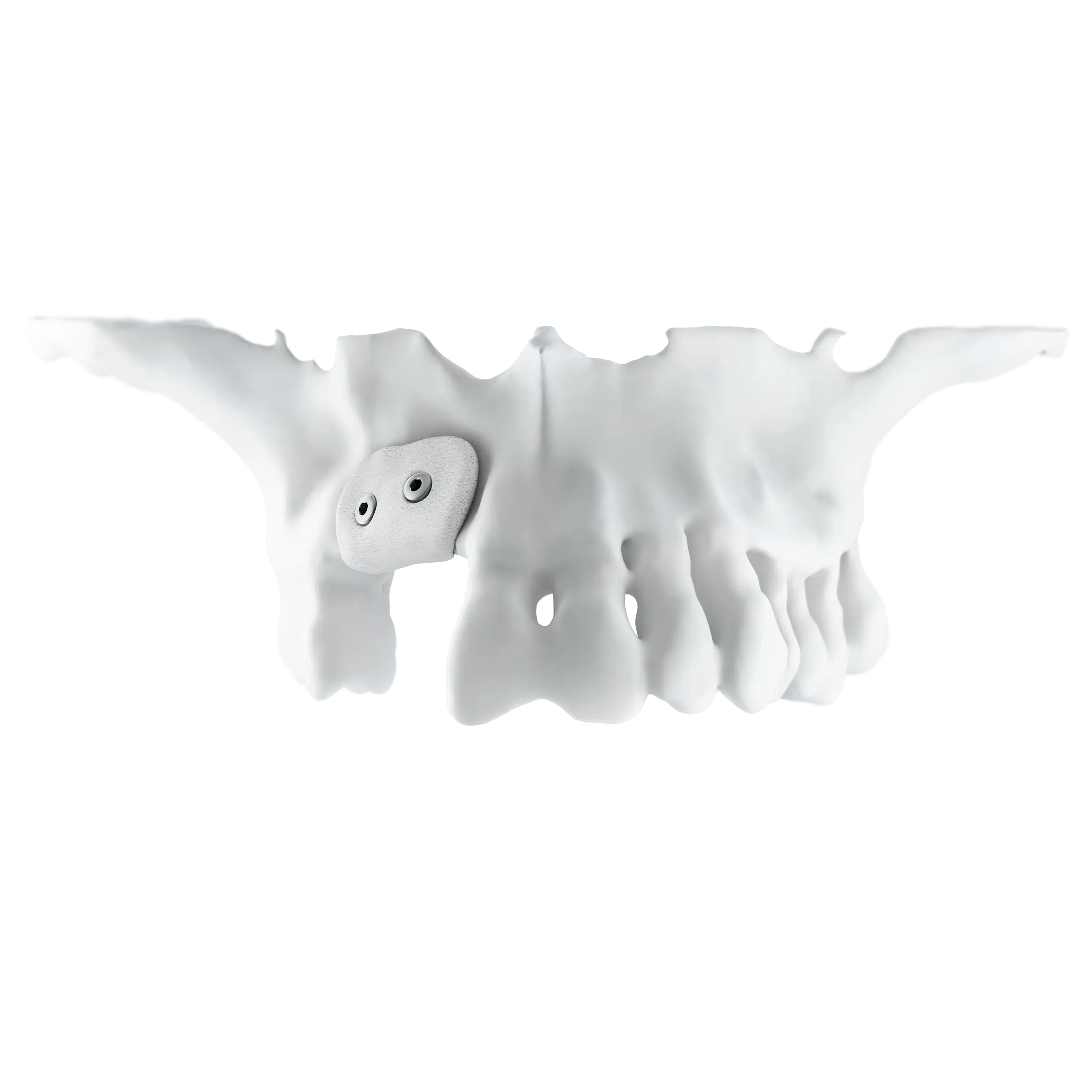
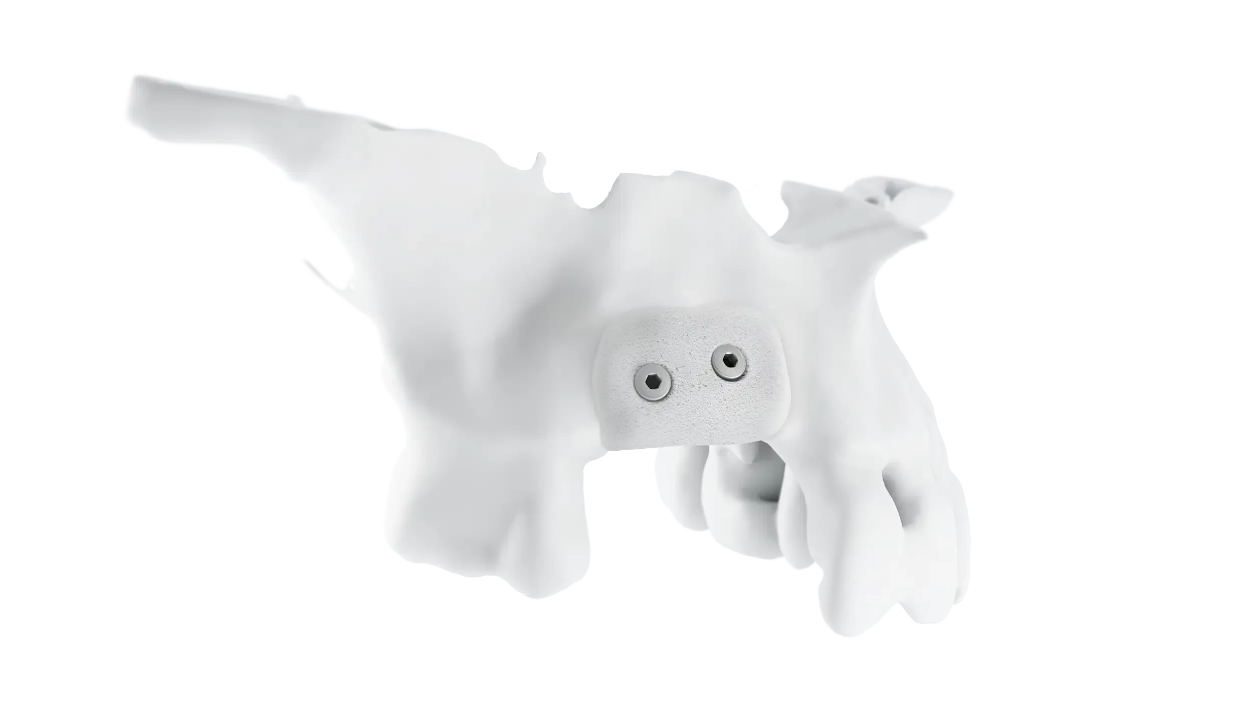
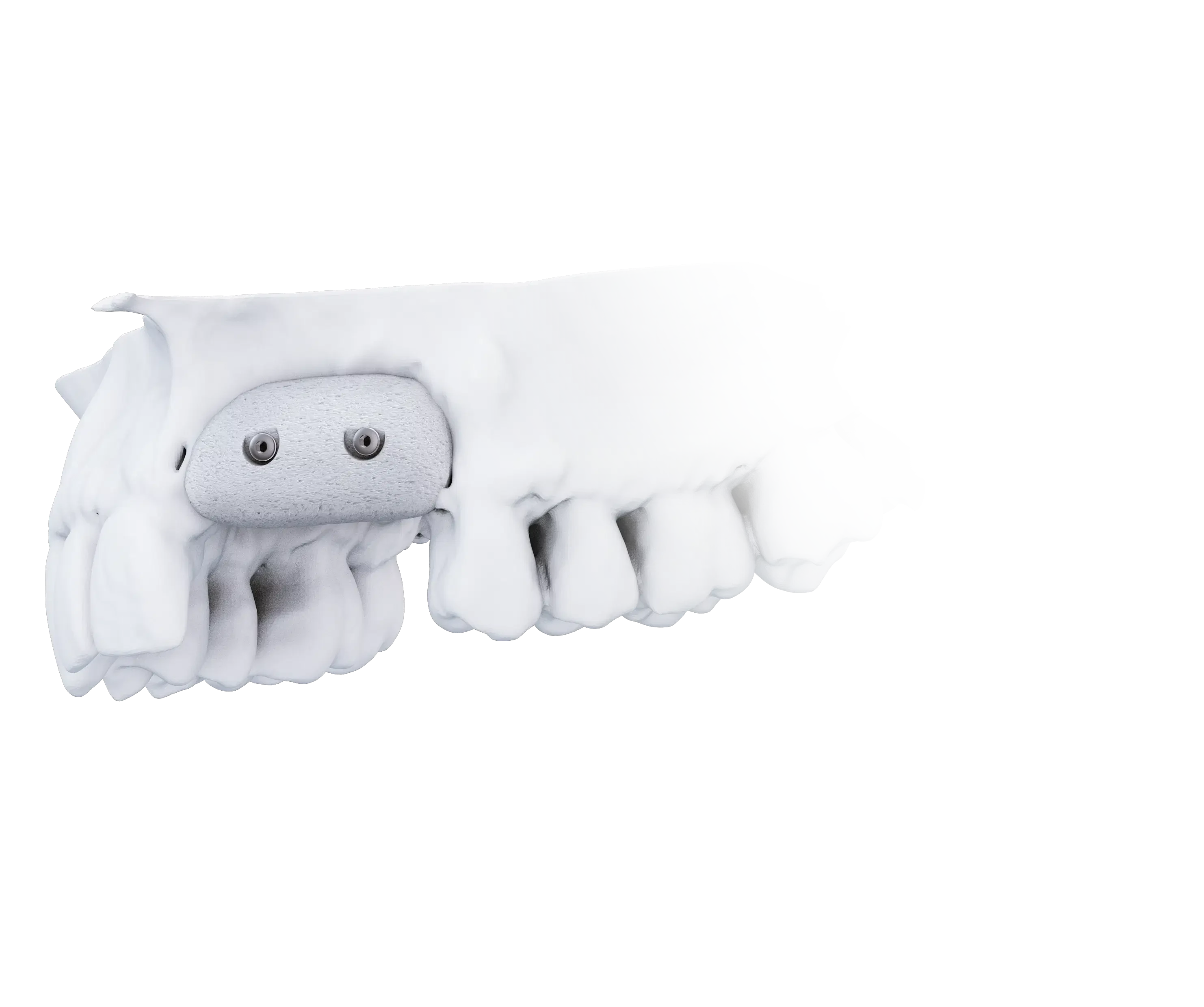
The Custom-Made Alternative to Anatomical Modelling
3D-BONE is a rapid, reliable solution for effective bone integration. Based on the patient's CBCT, the graft is produced using advanced CAD-CAM technology and subtractive multi-axis milling. It is ideal for small to medium-sized bone reconstructions, preparing the site for future implant placement.
Biological & Mechanical Advantages:
- Biphasic Composition: Composed of 30% slow-resorbing Hydroxyapatite (HA) to preserve bone volume, and 70% fast-resorbing Beta Tricalcium Phosphate (β-TCP) to facilitate rapid osseointegration.
- Optimal Porosity: Features a highly hydrophilic structure with 70% porosity (interconnected macro and micro pores) specifically devised to favour osteogenic processes.
- Mechanical Strength: Offers high resistance to mechanical stress during healing, boasting a compressive strength of 6 MPa and high dimensional stability.
- Surgical Efficiency: Eliminates the need for chairside anatomical modelling. Holes for cortical fixation screws are already present in the graft.
Indications for Use:
- Vertical regeneration
- Periodontal & peri-implant defects
- Sinus lifts & cystic cavities
- Post-extraction cavities & crestal defects
Available Graft Sizes
Depending on the extent of the defect, 3D-BONE is categorised into two primary dimensions:
- MINI: 10 x 15 x 15mm (for small reconstructions).
- MEDIUM: 24 x 22 x 15mm (for medium reconstructions).
- (Note: Every custom graft is supplied alongside a biocompatible PEEK replica to aid in surgical simulation and soft tissue management).
The 100% Digital Workflow
Diagnostic Acquisition
Acquire a Cone Beam CT (CBCT) of the jaw focusing on the defect, and upload the DICOM file to the BTK team via the web portal.
Digital Processing
The graft is designed using CAD modelling software to ensure the morphology, dimensions, and cortical screw holes perfectly fit the patient's anatomy.
Clinical Approval
You will receive a 3D digital model of the custom block. You can request remodelling or authorise production via a digital prescription.
Production & Delivery
The graft is milled from a synthetic bone block, while a PEEK replica is simultaneously produced. Both are rigorously decontaminated and packaged in a cleanroom, arriving ready for sterilisation.
Streamlined Surgical Procedure
- Simulation: Position the PEEK replica in the defect to verify fit and manage flaps.
- Preparation: Use the replica as a surgical guide to prepare the site for cortical screws.
- Fixation: Position the actual biomaterial graft and secure it firmly using cortical screws (we recommend using the BT SCREW Surgical Kit).